Digital and Modern Dentistry: How CBCT, Digital X-Rays, and Intraoral Scanning Improve Comfort and Results
Dental technology should make care easier to understand and more comfortable to receive. That is the guiding idea behind our use of modern tools such as CBCT 3D imaging, digital X-rays, intraoral cameras, and intraoral scanning. When used thoughtfully, these systems help us diagnose earlier, plan with precision, and deliver treatment that fits your bite and smile more naturally. Here is what each tool does and why it matters to you as a patient.
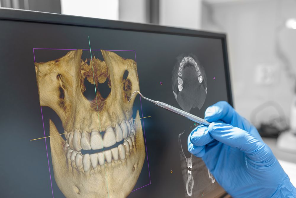
CBCT 3D imaging: detail when it counts
Cone beam computed tomography produces a three-dimensional map of your teeth, jaws, sinuses, and nerve pathways. Compared with traditional two-dimensional images, a CBCT scan can reveal hidden anatomy that influences decisions for dental implants, complex root canals, impacted teeth, or jaw joint concerns. We use CBCT selectively when the added information can change the plan or improve safety. The scan itself is quick, and your dentist reviews the images with you so you can see what we see.
Digital X-rays: clarity with prudent exposure
Digital sensors generate clear images instantly while using less radiation than traditional film. Because we follow an “as low as reasonably achievable” approach, X-rays are taken only when needed to answer clinical questions. That may mean bitewings to check for cavities between teeth, periapical images to evaluate root tips, or a panoramic view to examine wisdom teeth or jaw joints. Digital files are easy to compare over time, allowing us to monitor a small area rather than guessing.
Intraoral scanner: impression-free accuracy
An intraoral scanner uses a small wand to capture thousands of images and stitch them into a precise 3D model of your teeth and gums. The scan replaces messy impression materials for many procedures and makes it easier to design crowns, bridges, dentures, and implant restorations. Because the digital model is highly accurate, we can fine-tune your bite and contours for a comfortable feel and a natural look. Patients also appreciate seeing their scans on the screen, which brings planning conversations to life.
Intraoral camera and chairside visuals
High-definition images from a pen-sized camera make it simple to show tiny cracks, worn fillings, and early gum changes that are hard to see in a mirror. Clear visuals help you understand the “why” behind recommendations and allow you to participate in decisions with confidence.
Ultrasonic scaler: efficient, comfortable cleanings
During hygiene visits, an ultrasonic scaler vibrates gently to break up hardened tartar while flushing debris with a fine stream of water. The result is efficient plaque removal, smooth enamel, and fresher breath. Many patients find this method more comfortable than hand scaling alone, especially along the gumline.
How technology shapes your visit
- We start with your goals and health history, then select the right combination of images to answer specific questions.
- We review visuals together so you can see any problem areas and understand your options.
- We design a plan that fits your timeline and comfort preferences, using scans and photos to guide accuracy from start to finish.
- We follow up with maintenance tips and recall intervals tailored to your risk level for cavities or gum disease.
Benefits you can feel
- Earlier, more accurate diagnoses that prevent small problems from becoming big ones.
- Comfortable, efficient appointments with fewer remakes due to precise digital records.
- Thoughtful, case-by-case use of X-rays and scans to keep exposure sensible.
Thoughtful use, not tech for its own sake
Technology is most valuable when it answers a question or improves the outcome. We do not take images on a rigid schedule; instead, we choose the right tool for the right reason. This approach respects your time and health while supporting predictable, high-quality results.